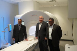
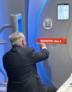
 The Food and Drug Administration (FDA) has provided clearance of a unique MRI whole body scanner that Roderic Pettigrew, Ph.D., M.D., Inaugural dean of the School of Engineering Medicine, envisioned in 2018. This scanner has 2.5 times the sensitivity for seeing small things that the most powerful prior whole-body scanners can and 5 times that of the most common scanners.
The groundbreaking Siemens Healthineers 3T Magnetom Cima.X is now commercially available for use and research. It is expected to detect life-threatening diseases early while allowing researchers to advance new life-saving treatments and preventative strategies. The scanner was designed and built after Dr. Pettigrew challenged the tech giant five years ago to “push the limits of MRI performance physics to the furthest point that could be safely used for the whole body.”
 “It is now a reality,” Dean Pettigrew said. “It will really improve our ability to identify disease early and prevent deleterious outcomes. We hope to identify vulnerable arterial plaque before it can cause a heart attack or stroke, identify breast cancer even earlier than we can today and minimize therapeutic intervention, or detect prostate cancer and pinpoint it before biopsy to enable prompt treatment or surgery without targeting the whole gland. This is a truly exciting and unique development in the field of whole-body MRI. It will also open doors for advancing research that we previously could not have imagined. As such, its potential to improve clinical care is just beginning to be explored”
Dr. Pettigrew, former and founding director of NIH’s National Institute for Biomedical Imaging and Bioengineering, worked closely with Siemens Healthineers for several years as the Cima was developed and produced.
Katie Grant, vice president of magnetic resonance at Siemens Healthineers North America, lauded the new scanner’s capabilities, saying it “possesses the strongest-ever gradient system for a clinically released whole-body MR scanner.”
“It can deliver penetrating new insights into oncologic, cardiac and neurodegenerative diseases,” she added.
The scanner works by using a powerful magnet with various electromagnetic coils, temporarily magnetizing the body, sending a radiofrequency pulse into the area being imaged to stimulate the hydrogen in tissue, then detecting the radiofrequency signal that the excited hydrogen nucleus returns.
 The signal is like those received by an FM radio, and a computer processes the signals to make images. These are used by clinicians throughout biomedicine including those in neuroscience, musculoskeletal, oncology and cardiovascular medicine. Most MRI scanners are only about 20% to 40% as strong as the Cima. The name Cima was chosen because this means “the highest point” or “summit” in Spanish. Its strong gradients can also make images of microstructures, like cardiac fibers, clearer. This makes it a powerful tool to help clinicians and scientists better understand and treat highly prevalent problems such as those that affect heart muscle structure and function. It can also measure water motion in tissue which is a sensitive marker of prostate cancer.
ENMED received the first scanner of its kind in May 2023.
Dr. Pettigrew’s work on the Cima project was sponsored by a Governor’s University Research Initiative (GURI) award. Established in 2015, GURI aims to attract transformative researchers to Texas universities who will bolster the standing of public universities and catalyze the Texas economy.

